"We should do an echo." In the GP's surgery this sentence often comes without any further explanation. The examination itself is harmless: no radiation, no preparation, and it takes about 30 to 45 minutes. What it delivers diagnostically is broader than most patients expect. What it does not deliver is narrower. This article shows you what a transthoracic echocardiogram measures, what it cannot see, how the appointment works in practice and when an echo on its own is enough.
What an echocardiogram measures and what it cannot see
Why an echocardiogram is done
In outpatient cardiology an echocardiogram has four main indications, which overlap in the consultation.
- Suspected heart failure. Breathlessness on exertion, swollen legs, sudden weight gain, orthopnoea (breathlessness when lying flat). TTE determines the left ventricular ejection fraction (LVEF) and classifies the type of heart failure (with reduced, mildly reduced or preserved ejection fraction) [Lang RM et al., JASE 2015].
- Suspected valve disease. A newly detected heart murmur, exercise intolerance, dizziness on standing up, syncope. TTE quantifies stenosis and regurgitation of the aortic, mitral, tricuspid and pulmonary valves and grades their severity [Galderisi M et al., Eur Heart J Cardiovasc Imaging 2017].
- End-organ assessment in the presence of cardiovascular risk factors. In long-standing high blood pressure, diabetes or a family history, TTE assesses left ventricular hypertrophy, diastolic function and atrial size. These findings change the individual risk and with it the treatment [Lang RM et al., JASE 2015].
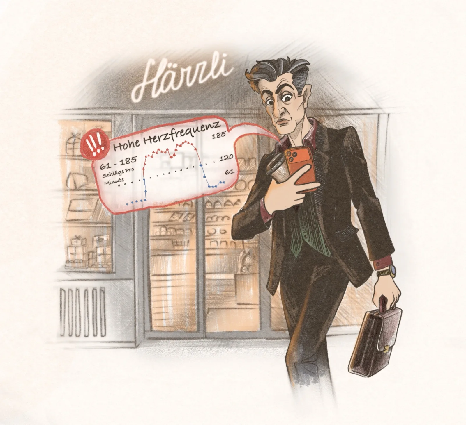
- Work-up after symptoms or findings. Atrial fibrillation flagged by an Apple Watch, unexplained chest pain, syncope, unusual tiredness, a previous stroke. TTE clarifies structural causes and provides the basis for further imaging or functional tests [Evangelista A et al., Eur J Echocardiogr 2008].
In our consultation we see the first two indications most often. The third is the least conspicuous and at the same time the most underestimated. Someone who has lived with high blood pressure for ten years is in a different risk class from a patient with the same blood pressure reading and no end-organ findings.
How the appointment works in practice
The examination is unspectacular. Here is the typical sequence in the consultation at Dein Team fürs Herz, from check-in to the written report.
- 0 to 15 minutes · History and clinical examination. Current symptoms, previous illnesses, medication, family history. Auscultation, blood pressure measurement, clinical status. These 10 to 15 minutes shape the targeted question put to the echo.
- 10 to 15 minutes · Preparation. You undress to the waist and lie on your left side. Three adhesive electrodes are attached for a simultaneous ECG. A water-soluble gel is applied to the ultrasound probe to improve sound transmission. It feels cool on application, the only unpleasant part. No special preparation on your part is needed. Eating, drinking and medication remain unchanged.
- 15 to 35 minutes · Image acquisition. The ultrasound probe is placed in several standard positions: parasternal, apical, subcostal and suprasternal. In each position two- and three-dimensional images, Doppler measurements and colour Doppler are recorded. You will be asked to hold your breath briefly or to shift position slightly. The machine records the loops digitally for later analysis.
- 35 to 50 minutes · Quantification and discussion of the findings. While you get dressed, the measurements are taken on the loops (volumes, ejection fraction, valve areas, pressure gradients). We then discuss the findings with you. You see your valves in motion, the pumping work of your ventricles, the relative size of your atria.
- Same day · Written report. The structured report follows the recommendation of the European Association of Cardiovascular Imaging on standardised reporting [Galderisi M et al., Eur Heart J Cardiovasc Imaging 2017]. It goes to your GP, and you keep a copy yourself.
Allow around 60 to 90 minutes in the consultation overall. The echo itself takes 30 to 45 minutes; the rest is history, ECG, frequently a bicycle exercise test (ergometry) and discussion of the findings. There is no radiation. There is no pain either. Bringing someone with you is always welcome, though never required.
What is measured in an echo
A complete TTE delivers far more than the famous "ejection fraction". The standard report covers around 30 quantitative parameters, which fall into four groups [Lang RM et al., JASE 2015; Galderisi M et al., Eur Heart J Cardiovasc Imaging 2017].
- Left ventricular function. Ejection fraction (LVEF, normal from 52% in men and from 54% in women), end-systolic and end-diastolic volume, wall thickness, mass, regional wall motion. With a reduced ejection fraction, the type of heart failure is defined [Lang RM et al., JASE 2015].
- Global longitudinal strain (GLS). A quantitative measurement of deformation along the long axis, more sensitive than LVEF for early impairment of systolic function. Normal value around minus 18 percent or more negative. Clinically useful in cardiotoxicity during chemotherapy, early cardiomyopathy and subclinical diabetic cardiomyopathy [Mor-Avi V et al., JASE 2011].
- Valve function. Aortic, mitral, tricuspid and pulmonary valves, each with quantification of stenosis and regurgitation via pressure gradients, valve areas and regurgitant volumes [Galderisi M et al., Eur Heart J Cardiovasc Imaging 2017]. Valve morphology is described as well (calcification, bicuspid aortic valve, mitral valve prolapse).
- Diastolic function and atrial size. E/A ratio, E/e-prime ratio, mitral annular velocity, left atrial volume index. These parameters are the axis for diagnosing heart failure with preserved ejection fraction (HFpEF) and for risk assessment in high blood pressure and atrial fibrillation [Lang RM et al., JASE 2015].
In addition, the pericardium (pericardial effusion?), the aortic root and the proximal aortic arch, the inferior vena cava (volume status), gross right ventricular function and, where relevant, intracardiac thrombi are assessed. The structured report is built on this data.
What echocardiography does not see
An honest answer to the most frequent question. There are diagnostic limits, and they are clearly defined.
- The coronary arteries themselves. TTE does not show whether there is a narrowing in a coronary artery and therefore a disturbance of blood flow to the heart muscle. It does, however, provide indirect clues through regional wall motion abnormalities, which may be caused by an insufficient blood supply to that area. [Lancellotti P et al., Eur Heart J Cardiovasc Imaging 2017]. Direct assessment of the coronary arteries requires a coronary CT (anatomical) or coronary angiography (anatomical and interventional in one).
- Microvascular disease. A dysfunction of the smallest coronary vessels escapes a resting TTE. It is investigated with stress echo, cardiac MRI or invasively with pressure and flow measurement during catheterisation. This question comes up frequently in women's heart health.
- Very subtle electrical abnormalities. Through the simultaneous ECG, TTE captures the rhythm during the examination, but no long-term profile. Atrial fibrillation that occurs only paroxysmally requires a long-term ECG or a wearable finding.
- Lung parenchyma and pleura in detail. Accompanying pleural thickening or pulmonary pathology is not systematically assessed in an echo. That requires a lung function test or lung imaging.
Echocardiography is the most commonly used modality for the evaluation of cardiac structure and function. Standardisation of measurements is essential to allow comparison across centres and over time.
These limits are part of the definition of the examination. A TTE is structural and functional imaging. Assessment of the coronary arteries is a separate examination with its own indications.
When an echo alone is enough and when it is supplemented
Diagnostic logic in cardiology is usually stepwise. TTE stands at the start of many pathways, and on its own it is sufficient for clearly defined questions.
In valve disease with a clear finding (for example moderate aortic stenosis without symptoms), TTE is sufficient as a regular follow-up examination. In heart failure with reduced ejection fraction, TTE provides the diagnosis, and follow-up measurements use the same method. In left ventricular hypertrophy as an end-organ finding of hypertension, TTE is the standard.
When the question goes beyond structural imaging, complementary tests are added. If coronary heart disease is suspected, a stress echo, a cardiac CT or coronary angiography follows [Lancellotti P et al., Eur Heart J Cardiovasc Imaging 2017]. In atrial fibrillation, a long-term ECG complements the rhythm assessment. In unexplained cardiomyopathy, a cardiac MRI provides the tissue characterisation that echo cannot capture.
In our consultation we discuss the findings in the same session. If complementary imaging becomes necessary, we schedule it directly. If no complementary imaging is needed, you hold the report in your hands the same day and go home with a clear plan.
Further reading in this cluster
These topics complement the cross-sections from this article:
- High blood pressure. When it is time to ask a cardiologist. Hypertension is the most common indication for end-organ assessment by echo.
- Heart check-up in Zürich. Costs and how insurance works. What a cardiological assessment with echo actually costs.
- Cardiac catheterisation. When it really becomes necessary. Anatomical assessment of the coronary arteries beyond the echo.
- Cardiac CT and calcium score in Zürich. Low-dose CT as a non-invasive alternative for direct coronary assessment.